
By Guy Eakin, PhD
In the below blog post, our Chief Science Officer, Guy Eakin, addresses a point of ongoing controversy in the Lipedema medical and research fields: Does Lipedema, as its name implies, actually involve edema, which is defined medically as swelling caused by fluid trapped in tissues of the body?
About 60% of Lipedema patients in the LF Registry tell us their thighs and calves swell. Indeed, LF’s founder and staff with Lipedema watch their legs change shape every day. Patients report that they feel their legs getting heavier or fuller as the day goes on, or that new subcutaneous fat appears on their legs, or that the skin on their legs feels “looser”.
At the level of the systems in the body, what is actually causing this swelling that Lipedema patients broadly report? This is an important question for several reasons.
Whether or not there is edema as defined in the medical literature has implications for how we understand the onset and progression of the disease, as well as which sorts of treatments may or may not work to alleviate symptoms. In some countries it is difficult to find enough therapists to treat swollen limbs, and some practitioners prioritize lymphedema patients (and de-prioritize Lipedema patients) for treatments like manual lymphatic drainage (MLD) on the belief that there is no edema in Lipedema.
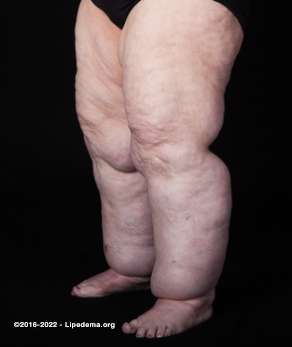
About 60% of Lipedema patients in the LF Registry tell us their thighs and calves swell.
It is also a timely question because some in the field are currently pushing to rename Lipedema on the basis of a disputed claim that there is no edema in Lipedema.
So we know from patient experience that swelling is a fundamental part of the experience of living with Lipedema. What does the rapidly evolving formal academic research tell us about what may be causing this swelling?
Dr. Eakin dove deeply into the published literature and talked to Lipedema researchers to better map our current scientific understanding of the role of edema, the lymphatic system, and comorbid conditions (especially lymphedema) in the onset and progression of Lipedema. The conclusion: As of now, there is currently evidence both for and against the implication of the lymphatic system and presence of edema, as classically defined, in Lipedema; and efforts to rename the condition are premature, since our knowledge about the disease continues to evolve.
If you're interested in the more technical discussion of this topic, check out Dr. Eakin’s blog below, or read the excellent open access review from the Crescenzi and Rutkowski laboratories.
Interested in going deeper, but need a quick crash course on the terminology? The lymphatic system sits alongside the blood system but runs one way. Tiny lymphatic capillaries in the skin sit beside tiny blood capillaries, feeding into increasingly larger vessels that ultimately end up in some organs in one’s core. The heart does not pump the lymphatics -- pumping is done by muscle contractions -- and there are one-way valves to prevent “backflow”. Any part of this system can break, and all are being studied by researchers. It remains to be seen which parts, if any, are involved in Lipedema. Are the teeny tiny capillaries “leaky”? Is pumping not working well because the valves are “finicky”? Or is the timing of pumping not efficient, resulting in insufficient clearance of fluid (which could explain why hands and feet are normal but the legs and arms are not)? These are all questions to which, hopefully, future research can provide some answers.
Historical perspective
The first papers to name Lipedema and describe it as a unique clinical condition considered edema, or retention of fluid, to be fundamental to the experience of women living with this condition. The choice of the term Lipedema, in their words, was to “describe large legs due to the subcutaneous deposition of fat in the buttocks and lower extremities and the accumulation of fluid in the legs” [1].
They differentiated Lipedema from many other edemas that were well known at the time, relating it more to a class of edemas that result from pressures exerted by outside factors, such as gravity when standing or inactive (orthostatic edema), or pressures exerted by a fetus on a woman’s blood and lymphatic vessels [2]. In doing so, they separated the origins of edema in Lipedema from the edema that occurs in familiar conditions such as lymphedema.
The mechanism by which they hypothesized that fluid might accumulate was logical and simple: “Because fat is a poor supporting structure, lacking firmness, it offers little resistance to the passage of fluid from the blood vessels into the tissue spaces; hence, the large limbs become even larger when patients are active on their feet” [1]. By this mechanism, they made no claim as to whether accumulation of fluid was due to failed lymphatic vessels.
Like all things biological, the simple answer often suffices in the early days, but becomes more complex as new data emerge. With nearly 80 years of progressively more sophisticated techniques, many investigators have tried to tease apart the relationship between adipose, fluid, and the symptoms experienced by women with Lipedema.
Debates in the field
The debate can be summarized as one between two “camps”. One claims that there is no edema in Lipedema. In this view Lipedema is defined solely as a characteristic accumulation of adipose, and any edema is considered to be a separate concern. In at least one often cited case, people with any edema were excluded from the study (they also removed people with a high waist-to-hip ratio from their Lipedema population, but that is a topic for another blog).
The other camp claims that not only is edema intrinsically connected to Lipedema, but the adipose and fluids work together synergistically in a manner that causes the Lipedema to worsen. Until recently, the data available to both camps had been relatively generalized views of entire limbs. These focused on examining lymphatic vessels in Lipedema using imaging techniques that allowed researchers to trace the lymphatic network. Chemical tracers were injected into or under the skin, and were recorded as they were cleared from the body. The results of these roughly 20 studies were equivocating, but can be separated by whether or not changes were seen in either the lymphatic structure (how the vessels looked), or function (how well did they work?).
Like all things science, one can pick apart these individual studies. And let’s be honest - being who we are - we’ve done that. Any study will have its limitations. We can ask whether the researchers watched the tracer long enough. Did they inject too little or inject too much? Did they include males as controls? Did they exclude lymphedema, as mentioned above, and thus eliminate any edema from their Lipedema population? It’s all there in the literature we’ve seen, and it’s all open to critique.
But what emerges are some general themes or nuances that should be considered. These are likely driven by a combination of professional needs and training. There is also a tension between developing clinically actionable data, and developing research data intended to support new therapy discovery through increasingly precise knowledge.
Nuance number 1 is the question of what defines “edema”. A clinical perspective might require evidence of fluid that is clinically significant, clearly affecting the patient, and possibly requiring intervention. A more technical perspective might have a lower threshold for the volume of fluid, and would suggest that any fluid at all is potentially noteworthy. This is regardless of whether it is clinically significant in a given patient.
Nuance number 2 is that the original definitions of Lipedema made no claim as to the origin of the accumulated fluid. In the first description of Lipedema, the excess fluid might come from capillaries, through the breakdown of membranes that divide groups of cells from one another, or from the dysfunction of the lymphatic vessels. Thus, nuance number 2 is that investigators focused only on lymphatics may not as easily see fluids that accumulate due to deficits in other tissues.
The modern era
The final nuance is made clear by two recent imaging studies. The first, a non-tracer MRI-based lymphangiography study by the Crescenzi lab at Vanderbilt University Medical Center, demonstrated a Lipedema-specific pattern of edema in the subcutaneous adipose tissue in a manner that is distinct from lymphedema or obesity controls [3].
The second study, from the Sevick Laboratory at the University of Houston, used a tracer injected at the skin and monitored in real-time as it was cleared. The results suggested increased pumping rates of and swelling of lymphatic vessels, and differences from typical structural changes seen in lymphedema [4]. These reports illuminate vagueness in earlier literature with respect to nuance number 1 (the definition of “edema”), but also remind us of a third nuance – the anatomical location of study. In these studies care was taken to differentiate any edema associated with the adipose beneath the skin from fluid being cleared from the skin itself.
These imaging approaches provide details effectively thought of as images taken at the scale of entire legs. While this broad coverage has many advantages, in recent years, more attention has been paid to carefully evaluating the microscopic structure of Lipedema-affected tissue, with techniques that precisely color-code different anatomic structures. Again, while early conclusions that Lipedema-affected tissue revealed enlarged blood vessels (e.g., reference [5]) were replicated in some more modern studies [6,7], other papers showed no remarkable differences between the structure of Lipedema vessels and controls [8,9]. Some papers included other measures that might imply leakiness, or permeability of vessels, included observation of infiltrating immune cells [6,7,8], though even this was not observed by all groups [9].
The future era
So what to make of this? What is the path forward? We reached out to a couple laboratories cited here, asking what they thought could be agreed between the camps. What would they personally be open to seeing that might resolve debate between these camps? And further, what would they need to be convinced of were someone to report data contrary to their findings?
Dr. Epameinondas Gousopoulos summed up the debate well, acknowledging additional nuances. The data we have so far is over merely correlative data. Resolution of the debate may require understanding whether any impairment of lymphatics can cause Lipedema-like consequences. Relatedly, could repair of damaged lymphatics alleviate Lipedema?
He’s right. But, Lipedema develops over decades in an individual, which makes this somewhat difficult to test currently. To determine whether lymphatic impairment can cause Lipedema-like consequences, rather than just being comorbid with Lipedema, would likely require reliable animal models of Lipedema, which have so far been difficult to create. The second issue, repair, may be more readily addressed, as many efforts to treat impaired lymphatics are currently underway. These might allow testing of whether Lipedema is alleviated when lymphatics are repaired, but may require longer observation periods than is customary for current lymphatics clinical trials.
Dr. Gousopoulos offers a variation on that idea and points also to the idea that lymphatic impairment would be easier to reconcile with Lipedema if the lymphatic problems could be found in the earliest stages of lipedema.
“Without doubt, data of lymphatic function would constitute here an integral part in answering the question raised above. Specifically, if the lymphatic compromise is mechanistically involved, this should be present already at the onset of the disease and not only after Lipedema is well-established. The functional impairment needs to address exactly this need and evaluate Lipedema stage 1 patients versus gender-, BMI- and anatomically-matched (lipohypertrophy) patients, preferably with lower BMI so that to exclude that state of being overweight or obese as a confounding factor.” - Dr. Gousopoulos (by email)
This, too, represents a reasonable approach, but one that may be difficult to test until an objective test for Lipedema, one that simplifies diagnosis at the earliest stages, becomes available.
Dr. Gousopoulos also brings attention, quite correctly, to a final idea that a narrow view of edema also avoids considering other cell types that might influence edema in Lipedema.
“The data currently available suggest that no gross morphologic [lymphatic] impairment underlies Lipedema, but, of course, the current state of research has yet not answered whether the micro-architecture shows any signs of compromise. At the same time, the involvement of other players such as distinct immune cell populations, well-known to interact with the vasculature and affect its function (e.g. permeability) cannot be disregarded. This complicates our understanding of a condition, about which we know admittedly less than we should.“ - Dr.Gousopoulos (by email)
Getting us to that future
Why do these debates matter? While seemingly academic, the near term goal of research is to provide a more objective diagnosis for Lipedema. Understanding edema’s relationship to Lipedema may help point researchers to ways of distinguishing Lipedema more readily from other conditions. Even if one pushes aside consideration of whether there is or is not edema due to Lipedema, we can all agree that many women are living with the consequences of both Lipedema and edema.
In the Lipedema Foundation Registry, 29% of women report comorbid lymphedema, and 56-70% report feeling of swelling or heaviness in their thighs [10]. In these women, edema and Lipedema may be connected in a vicious circle whereby accumulated lymph promotes proliferation of adipose [11], which itself further disrupts the lymphatic system. A more thorough understanding of the interplay between these two elements is needed in order to adequately treat these women.
Why would LF fund Lipedema research involve lymphatics if lymphatics haven’t been proven to be involved in disease progression? Two main reasons: Many patients report their legs swell and lymphatic-focused treatments help them, and even if lymphatics aren’t involved in disease progression, many diseases may eventually have treatments that are delivered to the lymphatics.
From a political standpoint, there are occasional calls to change the name of Lipedema. Some of the arguments in favor of a name change are based in a belief that edema is not an intrinsic component of Lipedema. Our view at the Lipedema Foundation is that this is not satisfactorily resolved, and a more clear understanding of the origins of Lipedema may suggest that fluid accumulation is indeed strongly associated with Lipedema.
We would be curious to hear your professional or personal perspective on the relationship between edema and Lipedema. Join this conversation on social media to discuss where you think the tools of research should be directed: Facebook | Instagram | LinkedIn | Twitter
CITED REFERENCES AND LINKS